Author: Nikita Rathod

Improving blood transfusion systems using an evidence-based approach

By Chris Bird, MSc Health Policy student at Imperial College and Project Manager in the System Engagement Programme at NICE

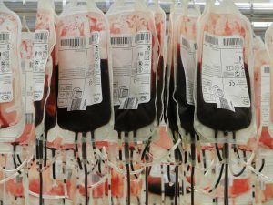
Today mark’s World Blood Donor Day – an event to celebrate and thank volunteers the world over, who generously donate blood to support life-saving care and to raise awareness of the continued need for donations of blood and blood products to support high quality safe care for patients who need it most.

(more…)

Towards safer and more equitable maternal health care

by Ana Luisa Neves, General Practitioner and Research Associate at Imperial NIHR PSTRC

Making motherhood safe is a human rights imperative. In the last 20 years, a steady decline has been observed in maternal mortality rates worldwide, but much more needs to be done: nearly 300,000 women still die every year because of pregnancy or childbirth-related complications (1). This means that a mother dies every two minutes.

(more…)

From Flatbush New York to London, via Paris: Ketosis-prone type 2 diabetes, a distinct form of diabetes we’re only beginning to understand

By Dr Shivani MisraHonorary Clinical Research Fellow, Faculty of MedicineDepartment of Medicine

SugarThis Diabetes Week, it’s important to remember that there are more than just two types of diabetes and how global insights into ethnic-specific types can benefit local people with diabetes.

(more…)

Mosquitoes, human health and environmental change

By Paul Huxley, Research Postgraduate, Faculty of Medicine, School of Public Health

Mosquito
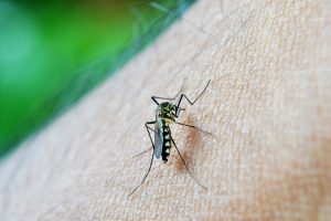
Ronald Ross, a British medical doctor of the late-19th and early 20th centuries, was first to identify the mosquito as the winged-insect carrier of malaria-causing parasites. Prior to this breakthrough, bad air (mal aria in Italian) was thought to have been the culprit. Together, Ross and Giovanni Grassi (who’s work, unlike Ross’, was controversially ignored by the Nobel Committee in 1902) uncovered a truth of huge ecological and epidemiological significance and sparked an ongoing international research effort aimed at answering fundamental questions about the processes that drive patterns of human morbidity and mortality caused by diseases carried by mosquitoes.

(more…)

Taking part in the UHC conversation

By Dr Ryan Li, Adviser, Imperial College London, Global Health and Development Group

Universal health coverage is about ensuring all people can get quality health services, where and when they need them, without suffering financial hardship. No one should have to choose between good health and other life necessities.

As part of World Health Day, Dr Ryan Li from the Global Health & Development Group who is an advisor for the International Decision Support Initiative (iDSI), which supports countries to get the best value for money from health spending, reflects on a visit to Vietnam and the principles for developing clinical quality standards in Low and Middle Income Countries (LMICs):

I remember very vividly two of the hospitals I visited in Vietnam, during my first field trip as a global health advisor for iDSI. In a central hospital in Hanoi, I saw an acute stroke centre that was spotlessly clean and gleaming with the latest equipment, with specialised stroke clinicians offering a range evidence-based treatments matching Western standards. In contrast, in a district hospital a mere two hours away from the capital city, I saw an elderly woman with suspected stroke who had been hospitalised for two weeks, seemingly not getting any better and not receiving any meaningful treatment (and there was no way to confirm the diagnosis as no brain imaging could be done). There was no question as to which hospital I would choose, if I could, if a relative or I were unfortunate to have a stroke.

The reality is that for most people, there is no choice – those who have the means to access the better hospitals, perhaps simply because they live closer to the city, likely get better treatment. This is unfair. Universal health coverage (UHC) is only truly universal if everyone has fair access to good quality health services, irrespective of where they live, what facilities they have access to, their education, income, religion or ethnic background.

Variation in quality is not a phenomenon unique to Vietnam, but is a reality in health systems across the world – even in relatively well-resourced and well-performing UHC systems such as the UK National Health Service. Some variation in quality may be acceptable, but one reason why unacceptable variation occurs is that there is a lack of clarity across the system about what is best practice.

Quality standards

In a bid to address this variation, the National Institute for Health and Care Excellence (NICE) in the UK introduced Quality Standards (QS): concise sets of statements that describe what is best practice in a given disease area (drawn from existing evidence based guidelines). For instance, what kinds of and how many antenatal checks a pregnant woman should ideally receive; sets out the practical steps required to achieve improvement; and most importantly quantifies the improvement. In essence, QS brings everyone together to identify the top five or 10 things that need improving nationally; and focus efforts towards raising standards in those areas.

Since 2012, iDSI has been working with India, China, Vietnam and Thailand to develop and implement QS as ways of tackling inequalities in healthcare quality; and to raise overall standards in key areas such as antenatal and maternal health, non-communicable diseases (stroke, hypertension, and diabetes) and antimicrobial resistance. We have drawn on our UK and international experience to create a guide to QS, which is now available in the resources section of the iDSI website and on our iDSI Knowledge Gateway.

Thailand, long seen as a success story of UHC, also recognises unacceptable variation in quality among public healthcare providers. In particular, there is now a push to raise and standardise quality in health promotion and disease prevention, beginning with QS in antenatal care. I was privileged to be invited as an international expert to observe and advise on this process. The discussions I heard among policymakers, clinicians and grassroots health volunteers were so rich that I can already anticipate insights and lessons that will go into the next version of the QS guide.

The enthusiasm and expertise of the stroke clinicians I met in the central hospital in Vietnam was unquestionable; the challenge is to sustain those excellent standards of practice and to ensure that all healthcare services across a country can reach those standards. iDSI’s vision is that everyone has fair access to quality healthcare, and we hope that our efforts in introducing and localising the QS model is a small step in the right direction.

Cholangiocarcinoma – the rare disease that’s on the increase

By Professor Simon Taylor-Robinson, Consultant Hepatologist and Professor of Translational Medicine at Imperial College London

Professor Simon Taylor-Robinson with colleagues

Cholangiocarcinoma is a rare primary malignancy arising from cholangiocytes, the endothelial lining of the biliary ducts, with an incidence 2500 cases of per annum in the UK. The only option for cure is surgical resection, but cholangiocarcinoma usually presents late when it grows sufficiently to block the drainage of bile from the liver, presenting with jaundice. By this point it is often irresectable, and palliative management includes holding open the ducts with stents to prevent blockage, and chemotherapy. One-year survival is only 5%.

The incidence of this insidious disease is increasing, and earlier diagnosis and better treatment are urgently required. Aetiological factors are thought to include exposure of the endothelium to carcinogenic compounds, and increased mutation induced by chronic inflammation, which may explain the association of chronic infection/gallstones, anatomical abnormalities, intrabiliary parasites, chronic biliary inflammatory disease. However, many patients do not have any identifiable risk factors.

The Lead Investigators in our research, as well myself, include Dr Shahid Khan, Honorary Clinical Senior Lecturer, Adjunct Reader from the Faculty of MedicineFaculty of Medicine Centre and Professor Richard Syms from the Faculty of EngineeringDepartment of Electrical and Electronic Engineering. Our work aims to tackle cholangiocarcinoma in five main areas:
                       
1) Novel biomarkers for cholangiocarcinoma. This takes advantage of the large numbers of patients with hepatobiliary malignancies including cholangiocarcinoma who are managed through the tertiary referral HPB services provided at Hammersmith Hospital, and utilises scientific expertise and infrastructure on both the Hammersmith and South Kensington sites. As part of a UKCRN-adopted programme we are establishing a biobank of body fluids from patients with various hepatobiliary malignancies in collaboration with other groups. We are applying metabonomic and proteomic analysis to blood, bile and urine samples to identify novel biomarkers for cholangiocarcinoma diagnosis and prognostication.

2) Genetic risk factors for cholangiocarcinoma. We have established a bank of DNA samples from patients with cholangiocarcinoma and with related diseases. DNA SNP analysis has demonstrated association of mutations in bilary transporters with development of cancer. Further analysis continues.

3) Epidemiology of Cholangiocarcinoma. It is known that the diagnosed incidence of cholangiocarcinoma is increasing. Work in our group in collaboration with Mireille Toledano (School of Public Health) has demonstrated the need for careful re-evaluation of these data as the real trend is obscured by changes in disease coding practice. Epidemiological work is continuing to identify associated factors which may give insight into cause, and assist in service planning.

4) Improving imaging in Cholangiocarcinoma. Current imaging techniques include ultrasound, MRI and CT scanning. However, optimising patient staging to accurately identify those who may benefit from resection or other therapies demands greater imaging resolution than these techniques can provide. In a collaborative project with the Richard Syms in the Engineering Faculty, we are
developing an MRI probe which could be placed inside the bile ducts to give fine detail on tumour anatomy and its breach of the ductal layers as it progresses. This has led to the development of novel coil technologies by our collaborating partners and offers exciting prospects for the future in this and other applications.

5) International and Domestic  Collaborations. In other countries infestation with biliary parasites is associated with a higher incidence of cholangiocarcinoma. We have previous, ongoing and evolving collaborations with Thailand, Egypt, and Bangladesh with the aim of comparing samples from fluke-related disease with our own cohort to identify differentiating factors. We also have collaboration with the Mayo Clinic and leading Norwegian researchers. Within the UK we have existing collaborations with Oxford and UCL, and are developing new relationships as part of the UKCRN scheme, with 6 new centres currently agreed

Ideabatic – where we are now…

By Kitty Liao and Abellona U of IdeabaticIGHI’s 2017 Student Challenges Competition winners

Kitty in the community where a vaccine campaign was being carried out

So much has happened since we won the Student Challenges Competition last year. The prize from the competition has been very helpful for us to secure our UK patent. Following that, we have recently submitted our global patent.

(more…)

Ending stigma and HIV transmission

By Dr Julia Makinde is a Research Associate with the International AIDS Vaccine Initiative at Imperial College London

It is estimated that there are 36.7 million people living with HIV globally with 1.8 million new infections in 2016 alone (1). This number represents an 11% drop in the number of new infections from 2010 . Some might consider this an achievement or a testament to the impact of strategic national and global policies aimed at tackling the epidemic. But in reality, these numbers mask the discrepant pace in the effort to tackle transmission and AIDS-related deaths in countries across the globe. UNAIDS recently reported a steep decline in new infections in sub-Saharan Africa against an alarming increase in the number of new infections in eastern Europe and central Asia in the same period (2). In the UK where the HIV burden is considerably lower, the number of new infections has been steadily falling (3). The reasons for these differences are complex and certain socio-economic factors have the potential to undermine the global effort to tackle transmission.

(more…)

Early Diagnosis of Lung Cancer: a Pathologist’s Perspective

By guest blogger, Dr Yu Zhi Zhang (Dennis), Clinical Research Fellow and Specialist Registrar in Histopathology at the National Heart and Lung Institute (NHLI), Imperial College London; on behalf of the National Centre for Mesothelioma Research (NCMR), National Heart and Lung Institute (NHLI), Imperial College London

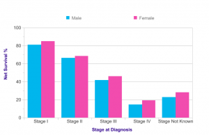
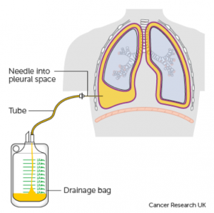
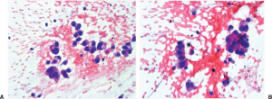
The 9th edition of the renowned Osler’s Textbook on the Principles and Practice of Medicine, published in 1921, dedicated only two (out of 1,139) pages to lung cancer, at which point the condition was described as “New Growths in the Lungs”. Almost a hundred years on, the patterns of epidemiology have shifted drastically, and lung cancer now is recognised as a major health problem globally with more than 1.8 million new cases diagnosed every year. It is the second most common cancer diagnosed in the UK for both men and women, with approximately 46,400 new cases in 2014. Unfortunately more than 60% of cases are diagnosed at Stage III and IV (Figure 1 (see below))[1], meaning the tumour has spread outside the lungs and into lymph nodes or other organs.

The prognosis of patients in these later stages is considerably worse than those diagnosed with Stage I or II disease where the tumour is confined in the lungs and is usually smaller. Stage III and IV disease also tend to develop significant symptoms preventing them from receiving aggressive treatments. It is estimated that more than 80% of patients with Stage I lung cancer will survive beyond 1 year, whilst less than 20% of those with Stage IV disease are expected to do so (Figure 2)[1]. This signifies the importance of early diagnosis, as it necessarily translates into treatment options with a curative intent.

A definitive diagnosis of lung cancer is usually made on tumour biopsies. Biopsies are small but representative portions of tissue from the tumour, obtained mainly via bronchoscopy (Figure 3) [2] and image- guided skin puncture (Figure 4) [3]. The tissue will then be processed and stained in the laboratory, before assessment under the microscope by a histopathologist. The minute features of the individual cells and overall architecture differentiate between cancer and normal tissue (Figure 5)[4]. If patients develop fluid around the lungs (also known as pleural effusion), a drain can be inserted (Figure 6)[5] and we can look for cancer cells floating in the fluid (Figure 7)[6].

Like all cancer types, the test results are discussed regularly at multidisciplinary meetings with doctors, surgeons and nurses where treatment plans are decided. Assessment of biopsies by a pathologist is a core component of the recently commissioned National Optimal Lung Cancer Pathway (NOLCP), which aims at improving early diagnosis of lung cancer in the UK.

Recent advances in lung cancer genetics have transformed our practice. The adoption of standard testing of biopsies with special stains for particular genes allows more refined diagnosis of lung cancer subtypes. Sequencing of relevant genes such as Epidermal Growth Factor Receptor (EGFR) reveal patients whose tumours are sensitive to targeted therapies. By assessing the expression levels of Programmed Death Receptor 1 Ligand (PD-L1), we are able predict a group of patients who will respond better to immunotherapies (Figure 8) [7], some of whom will receive long lasting disease control.

However, with such advances being undertaken at an unprecedented rate, many questions arise in the field of pathology. We are inevitably facing the scenario of “Doing More with Less”, i.e. there is a need to increase information yield with limited amount of clinical materials, which is often the case in initial diagnosis. Essential questions to be answered include: How do we ensure we have got sufficient clinical material for the ever-increasing number of ancillary tests? Can we provide the information in a timely fashion? How are we going to interpret and integrate the test results with clinical information? What is the relationship between genetic test results and what we see under the pathologists’ microscopes?

And perhaps more relevant to the theme: Is there a novel biomarker, test or modality that can lead to even more patients to be diagnosed earlier?

Hopefully these questions will be answered soon by translational research extensively supported by patient- derived tumour tissues. These tissue samples are either left over from biopsies taken for diagnosis, or are donated by the patients. For example, a major component of The 100,000 Genomes Project [8], funded by the Department of Health (DoH), is sequencing various cancer types on a massive scale, including many cases of lung cancer. The TRACERx study [9] looks at the genetic aspect of various parts of the same lung tumour, showing how cancer develops and changes over time.

Gone are the days when advances in the diagnosis and treatment of diseases were made primarily through observations by individuals. The research endeavours of the scientific community show strongly that there is now a greater need for multidisciplinary effort, active patient engagement, and international collaboration.

References

  1. Lung Cancer Statistics. Cancer Research UK. http://www.cancerresearchuk.org/health-professional/cancer-statistics/statistics-by-cancer-type/lung-cancer. Accessed 14/11/2017.
  2. Bronchoscopy under local anaesthetic. Cancer Research UK. http://www.cancerresearchuk.org/about-cancer/lung-cancer/getting-diagnosed/tests-diagnose/bronchoscopy-local-anaesthetic. Accessed 14/11/2017.
  3. Biopsy through the skin. Cancer Research UK. http://www.cancerresearchuk.org/about-cancer/lung-cancer/getting-diagnosed/tests-diagnose/biopsy-through-skin. Accessed 14/11/2017.
  4. Travis WD, Asamura H, Bankier AA, Beasley MB, Detterbeck F, Flieder DB, Goo JM, MacMahon H, Naidich D, Nicholson AG, Powell CA, Prokop M, Rami-Porta R, Rusch V, van Schil P, Yatabe Y; International Association for the Study of Lung Cancer Staging and Prognostic Factors Committee and Advisory Board Members. The IASLC Lung Cancer Staging Project: Proposals for Coding T Categories for Subsolid Nodules and Assessment of Tumor Size in Part-Solid Tumors in the Forthcoming Eighth Edition of the TNM Classification of Lung Cancer. J Thorac Oncol. 2016 Aug;11(8):1204-1223. doi: 10.1016/j.jtho.2016.03.025.
  5. Treatment for fluid on the lung (pleural effusion). Cancer Research UK. http://www.cancerresearchuk.org/about-cancer/coping/physically/breathing-problems/treatment/fluid-on-the-lung-treatment. Accessed 14/11/2017.
  6. Travis WD, Rekhtman N. Pathological diagnosis and classification of lung cancer in small biopsies and cytology: strategic management of tissue for molecular testing. Semin Respir Crit Care Med. 2011 Feb;32(1):22-31. doi: 10.1055/s-0031-1272866.
  7. Kerr KM, Tsao MS, Nicholson AG, Yatabe Y, Wistuba II, Hirsch FR; IASLC Pathology Committee. Programmed Death-Ligand 1 Immunohistochemistry in Lung Cancer: In what state is this art? J Thorac Oncol. 2015 Jul;10(7):985-9. doi: 10.1097/JTO.0000000000000526.
  8. The 100,000 Genomes Project. Genomics England. https://www.genomicsengland.co.uk/the-100000-genomes-project. Accessed 14/11/2017.
  9. Jamal-Hanjani M, Wilson GA, McGranahan N, Birkbak NJ, Watkins TBK, Veeriah S, Shafi S, Johnson DH, Mitter R, Rosenthal R, Salm M, Horswell S, Escudero M, Matthews N, Rowan A, Chambers T, Moore DA, Turajlic S, Xu H, Lee SM, Forster MD, Ahmad T, Hiley CT, Abbosh C, Falzon M, Borg E, Marafioti T, Lawrence D, Hayward M, Kolvekar S, Panagiotopoulos N, Janes SM, Thakrar R, Ahmed A, Blackhall F, Summers Y, Shah R, Joseph L, Quinn AM, Crosbie PA, Naidu B, Middleton G, Langman G, Trotter S, Nicolson M, Remmen H, Kerr K, Chetty M, Gomersall L, Fennell DA, Nakas A, Rathinam S, Anand G, Khan S, Russell P, Ezhil V, Ismail B, Irvin-Sellers M, Prakash V, Lester JF, Kornaszewska M, Attanoos R, Adams H, Davies H, Dentro S, Taniere P, O’Sullivan B, Lowe HL, Hartley JA, Iles N, Bell H, Ngai Y, Shaw JA, Herrero J, Szallasi Z, Schwarz RF, Stewart A, Quezada SA, Le Quesne J, Van Loo P, Dive C, Hackshaw A, Swanton C; TRACERx Consortium. Tracking the Evolution of Non-Small-Cell Lung Cancer. N Engl J Med. 2017 Jun 1;376(22):2109-2121. doi: 10.1056/NEJMoa1616288.


Figure 1: Proportion of Cases Diagnosed at Each Stage, All Ages (Source: Cancer Research UK)


Figure 2: One-Year Net Survival (%) by Stage, Adults Aged 15-99, England
(Source: Cancer Research UK)


Figure 3: A schematic representation of bronchoscopy
(Source: Cancer Research UK)


Figure 4: A schematic representation of image- guided skin puncture biopsy
(Source: Cancer Research UK)


Figure 5: Microscopic assessment of a lung adenocarcinoma
A. Low power; B. Medium power; C. High power. Figure adopted from Travis et al. [4].


Figure 6: A schematic representation of pleural effusion drainage
(Source: Cancer Research UK)


Figure 7: Individual tumour cells (Adenocarcinoma) from a patient with pleural effusion
A. Singly dispersed tumour cells; B. Tumour cells forming clusters. Figure adopted from Travis and Rekhtman [6].


Figure 8: Assessment of PD-L1 in lung adenocarcinoma
Strong staining (Brown) correlates with high level of PD-L1 expression in tumour cells. This predicts sensitivity towards PD-L1 based immunotherapy. Figure adopted from Kerr et al. [7].

Seek advice from a qualified healthcare professional before taking antibiotics

By Dr Timothy Rawson, Clinical Research Fellow, Esmita CharaniSenior Lead Pharmacist and Dr Enrique Castro Sanchez, Academic Research Nurse all from the Faculty of Medicine, Department of Medicine

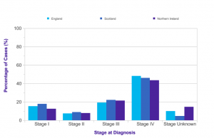
Antibiotics are a powerful resource that allows us to safely perform surgery, treat cancer with chemotherapy, and recover from infections that over 100 years ago would have killed even the fittest among us.

We are seeing however, a dramatic increase in infections with bacteria resistant to the killing effects of antibiotics (termed drug-resistant infections). These are antibiotics that until recently used to be effective. These resistant bacteria make many infections more and more difficult to treat – in some cases causing patients to die because we no longer have antibiotics that are able to manage the infection.

A major driver of drug-resistant infections is the overuse and misuse of antibiotics. We outline some of the key reasons for this below.

Firstly, some health problems cannot be treated with antibiotics, and it is important to know which ones they are. In some cases, health problems that can be treated with antibiotics need consideration about the right antibiotic for each person. For example, it would be important to consider whether people are taking other medications, whether they have got any other health problems, and even their preferences.

Talking to a health professional helps build relations that are proven to be essential for the experience of people related to antibiotic use. We now know that whilst patients are eager to receive more information about their use of antibiotics, previous experiences about having antibiotics prescribed (or not) gradually imprints expectations about antibiotics. Therefore, having the opportunity to discuss antibiotics with a healthcare professional may also have an impact on future attitudes and expectations when you feel unwell.

As all medications, antibiotics can have negative side effects that may be important to your current and future health. Often without previous discussion with a healthcare professional, it may be difficult to forewarn patients about such side effects and what to do should they appear. The ability to be able to warn someone about certain antibiotics may encourage them to keep taking them if they experience the side effect, or even ensure that they seek help immediately in rarer cases.

The conversation between healthcare professionals and patients about antibiotics are shaped by many factors that are not just related to health. These can reflect cultural beliefs and traditions, expectations, power relations between professionals and patients, the skills that the patient may have to deal with health problems, as well as social circumstances. Patients may feel compelled to access, or request antibiotics as a quick solution to a health problem that may prevent them from going to work. However, antibiotics are not always the solution and in some cases may worsen the problem due to the side effects that patients experience.

Conversations with healthcare professionals allows people to receive useful information about self-care measures that can resolve symptoms and prevent further health problems. For example, painkillers, fluids, and rest may be all that is required to deal with a cold. Being up to date on influenza vaccination, and washing hands thoroughly and correctly could ensure that the risk of catching infections is much reduced.

Studies show that whilst citizens are in agreement with healthcare professionals that the main reason causing drug-resistant infections is antibiotic use, they are not aware of the range of factors driving antibiotic use. Therefore, facilitating opportunities for these conversations to take place would be beneficial to raise awareness and optimise antibiotic use.

Encouraging citizens to engage with professionals in conversations about antibiotics would require that professionals embrace communications approaches and skills that result in meaningful and actionable information for patients and ultimately shared-decision making. Considering that the information related to infections is difficult to understand and process in the time allowed for typical consultations in primary care and due to the limited familiarity of the average citizen with scientific jargon/language, such conversations may be difficult. However, through the use of information aids  and a greater understanding of what information patients desire, we may be able to facilitate meaningful engagement with decision-making even in the most time pressured of situations.

In summary, we all have a role in using antibiotics more responsibly, through better communication approaches that encourage shared-decision making between citizens and healthcare professionals.