Blog posts

A retro approach to T cell expansion

By Faith Uwadiae

Edited by William Branchett

Recently we welcomed Professor Phillip Ashton-Rickardt for an insightful lecture and journal club discussion on his latest science paper, describing the identification of a novel protein important for T cell proliferation. (Read the full article here: https://www.sciencemag.org/content/348/6238/995.full)

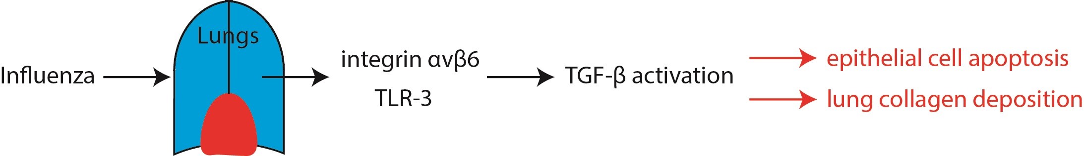
Effective CD8+ T cells responses are required for robust control of viral infections. However, during chronic infections, including clone 13 lymphocytic choriomenigitis virus (LCMV Cl13), the CD8+ T cell response becomes exhausted resulting in viral persistence. Ashton-Rickardt and colleagues used a forward genetic chemical mutagenesis approach to generate a panel of mouse lines, which they screened for enhanced CD8+ T cell responses during LCMV Cl13 infection. Screening more than 400 lines, the team identified three mutants with the desired phenotype, of which one, named Retro was the focus of this work. Retro mice benefited from increased CD8+ T cells proliferation and cytotoxic activity and almost complete clearance of virus. Interestingly, this improved CD8+ T cell activity in Retro mice was also sufficient to dramatically reduce tumour growth in mice given B16 melanoma cells.

The Retro mouse phenotype mapped to a single Alanine to Glycine mutation at position 1304 of an orphan gene which they named, lymphocyte expansion molecule (LEM). Retro T cells accumulated LEM as the mutation prohibited degradation of its mRNA. Contrastingly LEM heterozygous knockout mice had a 5-fold reduction in their CD8+ T cell numbers and higher LCMV Cl13 viral titre compared to wild type mice. Ectopic overexpression of human LEM resulted in increased human CD8+ T cells. Together this suggested that LEM was a positive regulator of CD8+ T cell proliferation.

Yeast 2 hybrid screening demonstrated LEM interacted with the mitochondrial protein, CR6-interacting factor-1 (CRIF-1), implicating it in oxidative phosphorylation. Indeed Retro mice had an increased rate of oxidative phosphorylation and LEM driven T cell expansion was found to be CRIF-1 dependent. Data from the lab of Erika Pearce (1) has previosuly suggested a link between mitochondrial reactive oxidative species (mROS) generated during oxidative phosphorylation and T cell proliferation. Fascinatingly, Retro mice had increased mROS levels and anti-oxidant administration prevented LEM induced CD8+ T cell expansion. Taken together, this suggests LEM plays an imLEM reduced
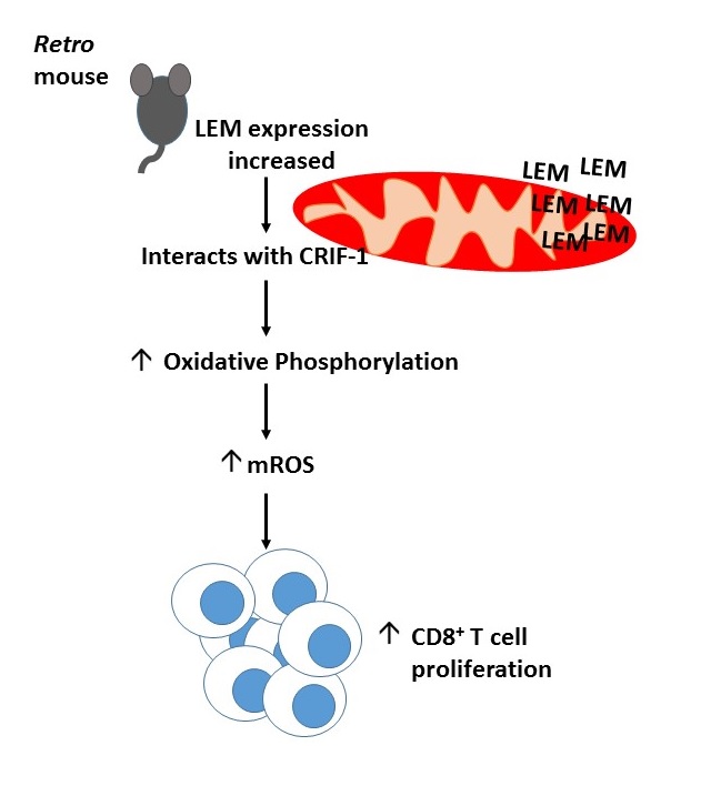
portant role in driving T cell proliferation by regulating mROS mediated oxidative phosphorylation.

After 6 long years of elegant molecule biology and dedication Ashton-Rickardt and colleagues have managed to identify a novel molecule with therapeutic potential for the treatment of cancer and chronic viral infection (summarised in the diagram). Much of the journal club discussion with Prof. Ashton-Rickardt focused on the methods of improving cytotoxic immunity by increasing LEM gene expression and the possible caveats of this in the clinic. There was also interest in the localisation of LEM within the mitochondria and the intricacies of mROS mediated T cell expansion. Professor Ashton-Rickardt was a clear advocate for the use of forward genetics to improve our understanding of key biological questions.

The team behind the paper have now formed a company called Immunar and are attempting to patent the therapeutic use of LEM with the hope of reaching the bedside within three years. We look forward to seeing how the story of LEM unfolds.

References

  1. Van der Windt et al, 2012, Mitochondrial Respiratory Capacity is a critical regulator of CD8+ T cell memory development, Immunity 36 (1):68-78

Interaction between S100A8/A9 with RAGE triggers a novel mechanism for NK cell activation

Written by Judith Secklehner

Edited by Faith Uwadiae

In our latest Journal Club session we discussed a recent publication in the Journal of Immunology. Narumi et al. investigated the role of S100A8/A9 (or Calprotectin) in a tumor environment and discovered a new role of this protein for NK cell activation via interaction with RAGE (receptor of advanced glycation end product).

The study focused on the heterodimeric protein calprotectin, which is formed of the two Ca2+-binding proteins S100A8 and S100A9. Calprotectin can be found in the cytosol of myeloid cells, but not in lymphocytes. Many intracellular and extracellular proinflammatory functions are attributed to calprotectin, essentially regulation of leukocyte adhesion and migration and promotion of cytokine production.  Although it can also act as an oxidant scavenger and possesses antimicrobial activity against bacteria and fungi.

There have been some inconsistent findings regarding the role of calprotectin for tumor promotion or progression. Evidence suggests that elevated levels of calprotectin can amplify inflammation and therefore promote tumor initiation in certain types of cancer.  However, some clinical studies indicate that in lung or gastric cancer increased levels of calprotectin were related with better prognosis for patients.

Therefore the aim of this work was to further investigate the in vivo role of S100A8/A9 in cancer. To this end murine pancreatic cancer cell lines were transfected with S100A8/9 cDNA and then inoculated subcutaneously in a syngenic mouse strain. Unexpectedly tumor growth was significantly supressed compared to controls inoculated with non-transfected cancer cells. This effect was also apparent in nude Balb/c mice, which lack thymus derived T-lymphocytes, therefore it was likely that NK cells were responsible for the tumor suppression.

Flow cytometry validated that an increased number of NK cells infiltrated the calprotectin-transfected tumor compared to non-transfected tumors. TLR4 and RAGE, are two pattern recognition receptors known to bind calprotectin. Whilst NK cells do not express TLR4, they were found to be positive for RAGE. To investigate whether RAGE/calprotectin binding was the mediator of the enhanced NK cell activation, antibodies to block RAGE were administered to the tumor-bearing mice. This a
bolished the ameliorating effects of RAGE/calprotectin signalling and increased tumor growth to similar levels observed in control cell lines. However RAGE/calprotectin binding alone did not seem NK RAGEto elucidate these effects but additional binding of NKG2D, an NK cell activating receptor, to an NKG2D-ligand was required. The authors concluded that RAGE/calprotectin has an enhancing or tuning effect on NKG2D induced NK cell activation.

This stimulating paper caused an animated discussion by our Journal Club group focussing on important criteria required for choosing suitable mouse strains and cell lines for a specific model.  We were intrigued by this novel finding and are awaiting the future work by Narumi et al, as they intend to investigate the role calprotectin production by neutrophils, for delivering NK cells activating signals to impede tumor progression.

Read the full article at: http://www.jimmunol.org/content/194/11/5539.full

CD8+ T cells follow neutrophil snail trails in virus-infected airways

Written by Caroline Anderson

Edited by John MacKey

The focus of our journal club this week was a recent paper from Science describing a novel mechanism by which neutrophils deposit chemokine-enriched way-markers to guide CD8+ T cells to the airways during influenza virus infection. (read the full article here http://www.sciencemag.org/content/349/6252/aaa4352.full).


snail-trail-best-jpg
Photo: Krstnn Hrmnsn, Creative Commons License

Whilst the role of chemokines in attracting effector immune cells to sites of immune insult is well established, how localized chemokine gradients are produced and maintained is incompletely understood. Likewise, the molecular mechanisms linking neutrophils and T cell recruitment in viral infections is unclear. Lim et al therefore set out to investigate the hypothesis that neutrophils are responsible for generating chemokine gradients that guide influenza-specific CD8+ T cells in the airways.

The authors first demonstrate that neutrophil depletion in a mouse model of influenza virus infection leads to impaired CD8+ T cell recruitment, localization and function in virus-infected trachea sections. Specifically they observed reduced numbers of both total and virus-specific CD8+ T cells, which lacked motility and remained distal to the epithelium. This dampened T cell response consequently led to a significant increase in viral load, demonstrating the importance of neutrophils in eliciting efficient T cell-mediated viral clearance. Using chemokine arrays and migration assays they were then able to identify CXCL12 as a key neutrophil-derived factor responsible for the recruitment of CD8+ T cells in this model. Importantly, the effects of neutrophil-depletion on CD8+ T cell recruitment were recapitulated using an antagonist against CXCR4, the chemokine receptor recognized by CXCL12. Striking immunofluorescence and scanning electron microscopy images revealed the generation of trails by crawling neutrophils, which were shown to contain CXCL12 and to persist in the trachea for up to 8 days post-viral infection. Finally, time-lapse in vivo two-photon microscopy images showed the deposition of CXCL12+ trails by neutrophils in the mouse ear, which appear to be followed by migrating CD8+ T cells. CXCL12 in neutrophil trails therefore appears to be critical for the effective recruitment and function of influenza-specific CD8+ T cells.

The journal club discussion centered on the exact timeline of neutrophil trail generation and the subsequent recruitment of the CD8+ T cells in vivo. It is clear that the neutrophil trails persist in the trachea for up to 8 days post-infection, yet it would be interesting to map trail deposition by individual neutrophils and to directly relate this to corresponding T cells over the course of the viral infection. In addition, we discussed the potential involvement of other chemokines and factors in this process, as well as the composition of the CXCL12-enriched deposits. We were also curious as to whether the same phenomenon occurred in response to influenza virus infection in other tissues, particularly in the lung, and in response to other viruses or inflammatory insults.

Influenza Promotes Collagen Deposition via αvβ6 Integrin-mediated Transforming Growth Factor β Activation

Journal club with Gisli Jenkins from 21/9/15

Written by David Salman, Richard Toshner and Joana Alçada

Edited by Sarah Allden

We recently welcomed Dr Gisli Jenkins, Clinical Associate Professor & Reader in Pulmonary Biology from the University of Nottingham to the IRD Journal Club.

His recent publication “Influenza Promotes Collagen Deposition via αvβ6 Integrin-mediated Transforming Growth Factor β Activation” served as a starting point to discuss various aspects of research in lung fibrosis. The paper explores the relationship between influenza and idiopathic pulmonary fibrosis via transforming growth factor β (TGF-β), a cytokine involved in development, inflammation and wound repair.

The authors hypothesised that influenza-induced epithelial cell damage would stimulate toll-like receptor 3 (TLR3) activity, a receptor that recognizes by-products from viruses such as influenza. This in turn may lead to activation of an epithelial specific cell surface molecule called integrin αvβ6, which would then turn on TGF-β signalling.

The results first indicate that influenza virus activates TGF-β in airway epithelial cells. Secondly, mutation of the epithelial cells with a non-functioning TLR3 protein inhibited this response, suggesting that the process is mediated through TLR3 signalling. Finally, blocking integrin αvβ6 with an antibody reduced influenza-induced epithelial cell death. Influenza was also shown to increase deposition of matrix materials, which are correlated with pulmonary fibrosis, throughout the lungs of infected mice. In addition, levels of integrin αvβ6 expression were increased in those mice. Conversely, blocking the expression of this integrin with antibody inhibited the development of a fibrosis-like picture in the lungs. Therefore, the Jenkins group have elegantly demonstrated a link between influenza infection and the mechanisms of pulmonary fibrosis development, through integrin αvβ6-mediated TGF- β and TLR3 signalling.

Print

The discussion Dr Jenkins kindly participated in was a wide ranging one, touching upon not just his own group’s research record, but also a more global view of translational research, with particular reference to idiopathic pulmonary fibrosis. He touched upon some of the molecules currently at the phase 2 trial stage for IPF, such as anti IL-13 and anti IL-4, as well as his thoughts upon the possible future directions that research in this area will take.

See Dr Jenkins’ full paper here http://www.jbc.org/content/289/51/35246.long

A novel role for Tregs in providing active TGF-β for immune regulation

Written by William Branchett

Edited by Judith Secklehner and Caroline Anderson 

The immune system has a huge capacity for destruction which, if unrestrained, can cause collateral damage to host tissue. Immune responses must therefore have ‘off’ switches to limit their magnitude and avoid inflammatory disease. Conversely, pathogens and tumours can hijack these mechanisms of immune regulation to favour their own survival. Understanding of immune regulation may therefore allow the design of drugs to manipulate overly exuberant or insufficient immune responses.

Transforming growth factor β (TGF-β) is a multi-functional cytokine that can dampen immune responses, in part by suppressing the pro-inflammatory activity of effector CD4+ T cells (Teffs). Previously, experiments from the labs of Fiona Powrie and Richard Flavell demonstrated that both regulatory T cells (Tregs) and TGF-β are required to prevent the pathogenic action of Teffs in a mouse model of colitis1. Unexpectedly, they also found that Tregs were not an essential source of TGF-β for this process1. So if not required to produce TGF-β, how do Tregs fit into the picture?

TGF-β is unusual among cytokines, as it must be released from a latent precursor complex to perform its biological functions. Among other mechanisms, latent TGF-β can be activated by interaction with certain integrins on immune and stromal cells. In their recent paper in Immunity, Worthington, Travis and colleagues highlight integrin-dependent TGF-β activation as a major role of Tregs in preventing T cell-driven inflammation2. The authors showed that integrin αVβ8 is highly expressed by Tregs in mice and humans, and allows Tregs to proficiently activate latent TGF-β. Expression of αVβ8 on Tregs was necessary to limit the severity of mouse models of colitis and skin inflammation, most likely by providing active TGF-β to pathogenic Teffs. Interestingly, mice lacking αVβ8 on their Tregs showed no signs of spontaneous inflammatory disease- suggesting that TGF-β activation by this mechanism is required during active inflammation but not at homeostasis.

There was much interest in this novel regulatory mechanism from the Journal Club group; particularly since production, rather than activation, of TGF-β is traditionally associated with the suppressive function of Tregs. As this story unfolds further, it will be exciting to hear more about the role of αVβ8 on Tregs in human disease and whether drugs targeting αVβ8 can selectively modulate TGF-β activity in the vicinity of inflammatory diseases and tumours.

TGFbeta Figure

Read the full paper here: http://www.cell.com/immunity/fulltext/S1074-7613%2815%2900172-7


References:

  1. Fahlén et al., Journal of Experimental Medicine. Vol. 201 no. 5 737-746
  2. Worthington et al., Immunity. Vol. 42 no. 5 903-915