Blog posts

Revolution rather than evolution? A journal club discussion on the use of gene editing based on the Netflix documentary series “Un-natural selection”

Written by Patricia Ogger

Edited by Max Kirtland


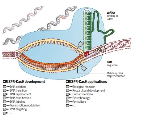
For millennia, humans have attempted to control nature to select traits in animals, change the environment, and eliminate infections and disease. In the last decade, the discovery and utilisation of the Clustered Regularly Interspaced Short Palindromic Repeats (CRISPR) system has provided us with a specific, efficient and cheap method to change nature instantly at the genetic level (see schematic for details – Doudna & Charpentier, 2014, Science).

The Cas9 enzyme generates breaks in double-stranded DNA next to a PAM sequence, matching the guide sequence (sgRNA) and thereby parts of the DNA can be deleted or added (Doudna & Charpentier, 2014, Science)

The explosion in the use of CRISPR, and the societal and environmental implications of this and other gene editing techniques were recently covered in the Netflix documentary “Un-natural selection”, by filmmakers Joe Egender and Leeor Kaufman. The four-part documentary series featured scientists, doctors, patients, conservationists and biohackers, showing their attempts and struggles to rewrite the code of life. 
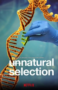
It further highlights some examples of applications such as treating rare diseases with specific genetic mutations; inducing gene drive to modify or eliminate wild animal populations to stop transmission of infections such as malaria and Lyme disease or eliminate rats; and the more controversial editing in human babies.  The series highlights some critical questions for us as scientists and our society, which we discussed in our journal club:


  1. What could and should gene editing be used for?
  2. Should these tools be available for everybody to use?
  3. What is a reasonable price for a live saving/life changing treatment?
  4. What might be the implications of induced gene drive?
  5. What ethical concerns does gene editing in human embryos raise?

After the discussion round, we concluded that gene-editing tools should be used for the research and development of new treatments for human diseases, though such applications would need to be tightly regulated. Challenges for this would be to identify diseases that could be treated by modifying a single gene in the patient and to make this affordable. Furthermore, these tools to modify somatic, non-reproductive body cells, should be only available for use in a safe and controlled environment, such as research institutes, to prevent the uncontrolled release of genetically modified organisms, which may have unforeseen consequences on the environment. We agreed that changes to human germline cells, like sperm and egg cells, should be prohibited as these changes would be inheritable and not only affect the individual, but also all their offspring. Therefore, genetically modifying human germline cells could have widespread consequences and would be uncontrollable. In animals however, a method called gene drive is already used to modify germline cells and thereby increase the probability of inheritance of a particular gene. By adding a specific gene to both alleles instead of the usual one, the probability of inheriting that gene increases dramatically, to a point where the whole population will carry that gene. For example, inducing gene drive might be a viable option to reduce transmission of diseases such as Malaria or Lyme disease, but such a drastic change to the mosquito and tick populations might have unforeseen long-term consequences for the ecosystem. It might therefore be safer and more beneficial to investigate other strategies to overcome these diseases in highly affected areas, such as developing better vaccines.

CRISPR may aid the development of complex therapies for rare diseases, as these can be very expensive due to a high chance of failure and small groups of patients, although public health care providers might still cover these expensive treatments. In the UK, the National Institute for Health and Care Excellence (NICE) releases guidelines on usage of therapies covered for certain diseases.

Overall, we concluded that while this documentary did not give a detailed explanation of how CRISPR actually works or a history of its discovery, it is a good foundation for discussion about gene editing and ethical concerns surrounding it. Providing a lay introduction to gene editing as a complex scientific and ethical issue, the series encourages a discussion about tomorrow’s gene editing technology that is ever increasingly on the horizon. We as scientists enjoyed the many different perspectives the series offers, some of which were very different from our own views and experiences.


Modulation of the gut microbiota in infants at high risk for asthma by Lactobacillus supplementation

By Rachele Invernizzi

The human gut is home to trillions of microorganisms that exert a marked influence on the host in homeostasis and disease. Research within the microbiome field is advancing at a rapid pace; recent work has shown that the composition and function of the gut microbiota are symbiotically linked with host health, and altered in several immune-mediated disorders, including asthma. With the intention of modulating the intestinal microbiota, early probiotic administration has been used for the prevention of allergic diseases in infants, with variable success.

Durack and colleagues (1) wanted to explore the maturation of gut microbiota over the first year of life in infants at high risk for asthma, and whether it was modifiable by early-life Lactobacillus rhamnosus GG (LGG) supplementation. They found that early-life gut microbial development was distinct from seen adults but malleable, and infants at high risk of asthma were therefore responsive to LGG supplementation. This meant a daily oral dose of probiotic temporarily modified the gut microbiota of these infants, changing the bacterial composition and metabolic profile of the gut while altering the immune system by potentially allowing more regulatory T cell development.

This study reinforces the observation that the gut microbiome is altered and has delays in diversification in infants at increased risk of childhood asthma. More importantly perhaps, given the difficulties in making significant, disease changing alterations to the gut microbiota in adults, they show that early-life strategies such as probiotics could prove effective.


(1) Durack et al, Delayed gut microbiota development in high-risk for asthma infants is temporarily modifiable by Lactobacillus Nature Communications. (2018). 9:707. https://www.nature.com/articles/s41467-018-03157-4

The gut microbiome: a trigger for asthma in childhood?

By Rachele Invernizzi

The human microbiome is composed of approximately 100 trillion bacteria and our intestinal tract is where the majority of these bacteria reside. The gut microbiota and their metabolic products exist in a dynamic state, which varies throughout the lifetime of the individual. This is particularly true during the first 18 months of life as the gastrointestinal tract becomes colonised and communities of bacteria form in various niches throughout the gut. There is a growing appreciation that the gut microbiota plays a vital role in host physiology as well as in the pathophysiology of several disorders, including asthma.

The primary objective of this study (1) was to phenotype the gut colonisation patterns during the first year of life and the associations of these patterns with the later risk of asthma in a prospective cohort of 700 children. The authors looked at the abundance and diversity of the bacterial species in the children’s guts and how they changed from the first week to the first year of life. They found that the state of the gut microbiome is associated with diagnosis of asthma at age 5, but fascinatingly these observations are only apparent in children born to asthmatic mothers.

Taken together these findings highlight the importance of the maturation of the gut microbiome during the first year of life in the development of asthma, especially in children born to asthmatic mothers. This raises important questions about the nature of disease inheritance. Are children of asthmatic parents more susceptible solely as a result of genetics, or also because of imbalances in microbiota they inherit from their mothers? Would correcting this reduce the risk of developing asthma? And how do these resident gut microbes influence events in the lungs?

  1. Stokholm et al, Maturation of the gut microbiome and the risk of asthma in childhood. Nature Communications. (2018). 9:141. https://www.nature.com/articles/s41467-017-02573-2

Type 2 innate lymphocytes guard against acute lung injury

Written by Chloe Pyle

Edited by Lauren Headley


Idiopathic pulmonary fibrosis (IPF) is a progressive lung disease of unknown cause, which results in hardening of the lung parenchyma due to collagen deposition and loss of pulmonary function. The role of the immune system in lung fibrosis is not well defined, although previous studies have shown that immune suppression is associated with decreased survival in acute exacerbations of IPF. The new study we discussed (1) addresses the contribution of population of immune cells, those that express the molecule ICOS, on the outcome of acute lung injury in a mouse model of lung injury and fibrosis.

ICOS-deficient mice were significantly more susceptible to fibrotic lung injury, resulting in severe weight loss, vascular leakage and airway oedema, but collagen deposition was unaffected. Type 2 innate lymphoid cells (ILC2s) were the first ICOS+ population to expand upon injury, and the authors speculate that the IL-5 produced by these cells is key in protecting from acute lung injury. In the lungs of patients with IPF these cells were deficient when compared to the lungs of healthy individuals. Their data suggests that lung resident ILC2s are poised to respond rapidly to injury and play a key role in regulating vascular permeability, which may confer protection during acute lung injury whilst not affecting the fibrotic process (see cartoon).


  1. ICOS protects against mortality from acute lung injury through activation of IL-5+ ILC2s. Hrusch CL, Manns ST, Bryazka D,…. Sperling AI. Mucosal Immunology (2018) 11:61-70. https://www.nature.com/articles/mi201742

TLR4 localization: Shaping inflammation is a battle of the lineages

By Dhiren Patel

Edited by Chloe Pyle and Faith Uwadiae


In the recent ‘Compare & Contrast’ journal club, we had the pleasure of discussing two excellent papers based on the immunopathological roles of Toll-like Receptor 4 (TLR4). The two papers presented the importance of the localisation of TLR4 on stromal or haematopoietic cell types and their contribution in inducing pathology in different airway disease models.

Toll-like receptor 4, is a pattern-recognition receptor that is expressed on array of cells and a common agonist for this receptor is Lipopolysaccharide (LPS), a sugar molecule highly expressed on gram negative bacterial cells1. The importance of TLR4 expression on immune cells such as granulocytes, neutrophils and eosinophils in driving an immune response against various pathogens has been published on extensively. Additionally, structural cells such as epithelial and endothelial cells have been shown to express TLR42. In many cases however, dysregulation of cell signalling from both haematopoietic and stromal compartments can lead to over-exuberant inflammation and subsequent collateral tissue damage and remodelling. TLR4 signalling is speculated to be important in contributing to hyper-inflammatory situations but whether this due to a direct effect of TLR4 expression on effector immune cells or an indirect effect of TLR4 signalling within stromal cells, is something that was addressed within the discussed papers.

Paper 1

The first paper was ‘House dust mite allergen induces asthma via Toll-like receptor 4 triggering of airway structural cells’, by Hammad, H et al., published in 20093. This paper utilised irradiation techniques to eliminate host immune cells and leave structural cells such as airway epithelial cells intact within irradiated mice. They also performed bone marrow transfer experiments from respective Wild Type and TLR4 -/- mice to irradiated animals to produce mice that in turn either lack TLR4 expression on epithelial cells (Wild Type mouse bone marrow transferred to an irradiated TLR4 -/- animal) and mice that lack TLR4 expression on immune cells (Wild Type mouse bone marrow transferred to an irradiated TLR4 -/- mouse). They used an LPS model of inflammation and a House Dust Mite (HDM) model of allergic airway disease to determine which of the examined cell types expressing TLR4 was critical in driving immunopathology. Both of these studies displayed that TLR4 expression on radioresistant airway epithelial cells was critical in driving both disease types.


In the LPS model, the authors observed elevated neutrophil, monocyte and dendritic cell infiltration in mice with TLR4 expression in the epithelium. They then went on to display that TLR4 expression on epithelial cells was important in dendritic cell maturation which in turn aids the development of LPS-induced inflammation. In the House Dust Mite model of allergic airway disease, mice with intact TLR4 expression in epithelium developed elevated airway infiltration of key effector cells such as neutrophils, eosinophils and lymphocytes to a similar extent to that of Wild Type mice administered HDM. Additionally, upon administration of an intratracheally-delivered TLR4 antagonist (assumed to recapitulate blocking TLR4 expression specifically on epithelial cells), cellular recruitment and cytokine production was ablated to similar levels as globally deficient TLR4 -/- mice and mice lacking TLR4 expression within the immune compartment.

Paper 2

The second paper discussed was ‘Distinct Tlr4-expressing cell compartments control neutrophilic and eosinophilic airway inflammation’,by McAlees, J.W. et al., published in 20154. This paper intriguingly concluded opposing findings to those observed in the Hammad paper. This paper avoided the utilisation of irradiation techniques and exploited genetic strategies to manipulate TLR4 expression on respective immune or structural cell compartments. TLR4fl/fl  mice were crossed with Vav1-Cre mice to inactivate TLR4 on immune cell types. Additionally, TLR4fl/fl  mice were crossed with Shh-Cre mice to remove TLR4 expression on epithelial cells.

Upon administration of the TLR4 agonist LPS, TLR4fl/fl-Vav1-Cre mice had reductions in neutrophil recruitment, chemokines and other inflammatory parameters compared to respective TLR4fl/fl-Shh-Cre mice. This indicates that it is indeed TLR4 expression in the haematopoietic cell compartment which drives this immunophenotype. Similar to the previous publication, a House Dust Mite sensitisation and challenge model was exploited. Intriguingly the TLR4fl/fl-Vav1-Cre mice had reductions in neutrophils numbers but eosinophilic infiltration in the airways was unchanged, however eosinophil recruitment was ablated in TLR4fl/fl-Shh-Cre suggesting that TLR4 expression in haematopoietic cells is important in driving neutrophilic inflammation but TLR4 expression on the epithelium is critical in inducing eosinophilic inflammation in allergic airway disease. Similar findings to the genetic studies were recapitulated when the authors employed irradiation studies similar to that of Hammad et al. Although epithelial expression of TLR4 does indeed have an important role in driving eosinophil recruitment, the study found that immune cell expression of TLR4 was critical in driving the pathology in their respective LPS and HDM models of inflammation.


journal-club-figure-dhirenTwo papers with such contrasting conclusions led to extensive discussions within the group. There was a general inclination towards the recent McAlees paper, and the major reasoning behind this was primarily linked to the irradiation techniques employed. The major argument was the effect of radiation on the inflammatory status of structural cells post-radiation treatment and additionally bystander cells such as alveolar macrophages that may be left intact. Additionally bone marrow transfer techniques have also been linked with inducing inflammation alone. On the other hand, despite employing genetic manipulation, doubts were also raised about the specificity of compartmental deletion of TLR4 in the respective TLR4fl/fl-Vav1-Cre and TLR4fl/fl-Shh-Cre mice.

Overall it was agreed that whilst impressive and inventive techniques were used to observe compartmental contributions to the pathology of a disease, there still is a need to develop better models to deduce such hypotheses.

  1. Hoshino, K. et al. Cutting edge: Toll-like receptor 4 (TLR4)-deficient mice are hyporesponsive to lipopolysaccharide: evidence for TLR4 as the Lps gene product. J. Immunol. 162, 3749–52 (1999).
  2. Saito, T., Yamamoto, T., Kazawa, T., Gejyo, H. & Naito, M. Expression of toll-like receptor 2 and 4 in lipopolysaccharide-induced lung injury in mouse. Cell Tissue Res. 321, 75–88 (2005).
  3. Hammad, H. et al. House dust mite allergen induces asthma via Toll-like receptor 4 triggering of airway structural cells. Nat. Med. 15, 410–6 (2009).
  4. McAlees, J. W. et al. Distinct Tlr4-expressing cell compartments control neutrophilic and eosinophilic airway inflammation. Mucosal Immunol 8, 863–73 (2015).

Chemokine cascades drive the education of metastasis-promoting macrophages

Written by Caroline Anderson

Edited by William Foster and Arunon Sivananthan

This week we were fortunate enough to welcome a world-renowned biomedical scientist to the IRD journal club. Professor Jeffrey Pollard is currently the Director of the Medical Research Council Centre for Reproductive Health at the University of Edinburgh. His seminar earlier in the day had focussed on the role of the tumour microenvironment, and metastasis-associated macrophages (MAMs) in particular, in breast cancer metastasis. Much of the work he discussed can be found here: Kitamura et al, JEM, 2015 (PMID: 26056232). Pollard Figure

Both pro- and anti-tumorigenic immune cells are found in association with tumours. Indeed, it appears that tumour-associated macrophages can be influenced by the tumour microenvironment into adopting a more tumour-promoting phenotype (Kitamura, Qian and Pollard, Nature Reviews Immunology, 2015). Previous work showed that antibody blockade of CCL2 signalling inhibited the recruitment of inflammatory monocytes (IMs) and reduced MAM numbers in lungs with metastatic tumours (Qian et al, 2009). As such, Kitamura et al set out to test the hypothesis that the CCL2-CCR2 axis was involved in regulating MAM function and MAM-mediated metastasis of cancer cells.


Firstly, they performed a microarray to identify potential downstream targets of CCR2 signalling in MAMs. Interestingly, Ccl3 expression was significantly downregulated in macrophages lacking CCR2 when compared to wild-type macrophages. Secretion of CCL3 increased when wild-type but not CCR2-deficient macrophages were stimulated with recombinant CCL2. Hence, CCL2 signalling via CCR2 can increase CCL3 secretion from macrophages. This chemokine-induced chemokine secretion was also apparent when human monocyte-derived macrophages were stimulated with recombinant human CCL2 or conditioned medium from human breast cancer cells, whilst the effect was abolished in the presence of anti-CCL2.


The Pollard lab make extensive use of a genetically engineered mouse model of breast cancer called the Polyoma Middle T mouse model. In this model, the Polyoma Middle T oncogene is expressed specifically in the mammary epithelium, leading to spontaneous metastases which preferentially migrate to the lung. Using this model, Kitamura et al show that genetic deletion of CCL3 or CCR1 in macrophages caused a decrease in the number of metastatic foci and reduced MAM numbers in the lung. Conversely, adoptive transfer of CCR3+ IMs to Ccl3-deficient mice increased metastatic seeding of cancer cells.


The CCL3 receptors, CCR1 and CCR5, are predominantly expressed by macrophages rather than breast cancer or endothelial cells, suggesting that CCL3 acts in an autocrine manner on MAMs. Using CMFDA-labelled inflammatory monocytes, the authors found that CCR1 deficiency affected the retention of MAMs in the metastatic lung but not the recruitment or differentiation of IMs to the lung. Finally, the interaction between tumour cells and MAMs was found to be dependent on a4-integrin expression by leukocytes and expression of its ligand, VCAM-1 by breast cancer cells. Taken together, the paper concludes that a CCL2-induced signalling cascade in macrophages leads to enhanced metastatic seeding of breast cancer cells through the increased retention of MAMs.


The journal club discussion centred around Professor Pollard’s experiences of working in the US and the opportunities afforded by working abroad. Moving back to science, we touched on the intriguing evidence that chemokines play roles above and beyond chemotaxis, which could have consequences for the therapeutic targeting of these signalling molecules in disease.


Hold your nerve; a new way to halt MPN progression

Written by William Foster

Edited by Judith Secklehner

On Monday the 15th of February the NHLI welcomed Dr Simón Méndez-Ferrer for a lunchtime seminar. Dr Méndez-Ferrer has recently moved from Madrid to Cambridge, where he has established a group in the Haematology department. Dr Méndez-Ferrer studies the multisystem regulation of the haematopoietic stem cell niche and his work has clinical implications for bone marrow transplantation procedures, as well as insight into specific forms of leukaemia. In Simón’s talk he discussed haematopoietic stem cell trafficking; paying attention to the importance of microenvironments; with focus on the importance of neural signal modulation within the bone marrow and how Mesenchymal Stem cells(MSCs) regulate inflammatory cell trafficking.

The focus of the journal club was on a paper published by the Méndez-Ferrer group in Nature in 2014. The paper is titled “Neuropathy of haematopoietic stem cell niche is essential for myeloproliferative neoplasms” and can be found at http://www.nature.com/nature/journal/vaop/ncurrent/full/nature13383.html

Myeloproliferative neoplasms (MPNs) are a family of diseases characterised by the bone marrow overproducing one or more blood cell types. The disease manifests as mutations in the Haematopoietic stem cell (HSC) compartment.  The majority of MPN patients show a common mutation in Janus Kinase 2 (JAK2) in their HSCs. This mutation causes constitutive activation of the kinase, leading to haematopoietic cells becoming more sensitive to growth factors like thrombopoietin, which leads to over-proliferation.

Simón’s previous work showed that the HSC niche is controlled by nestin+ MSCs which are innervated by the sympathetic nervous system. This paper demonstrates that during MPN pathogenesis, the MSC/HSC cellular circuit is corrupted. In both human patients and mouse models nestin+ MSCs, sympathetic nerve fibres and their supporting Schwann cells were depleted. In pathological conditions, loss of stem cell populations can often be attributed to loss of their quiescence, leading to differentiation away from the stem cell phenotype. However in this work, MSC depletion was shown to be due to an increased rate of apoptosis. Through the use of multiplex ELISA assays, the group showed that IL-1β is upregulated by mutant HSCs, and this can sensitise the neural cells for apoptose. Time course experiments showed that MSC apoptosis occurs after the loss of innervating nerve fibres, and so the paper then goes on to show that if the nerve cells are protected, so are the MSCs.

Experiments in the second half of the paper aim to prevent the development of MPN pathology through the use of neuroprotective agents (4-methylcatechol) and β-3 adrenergic agonists (BRL37344). The use of β-3 adrenergic agonists supplement the MSCs which are not receiving stimulus from the sympathetic nervous system, and BRL37344 treatment prevented the neutrophilia and thrombocytosis seen in unchecked MPN disease. The paper also displays some impressive histological work, showing that use of BRL37344 prevents the build-up of fibroblastic and bone tissue deposits. In addition to initial prevention of disease manifestation, BRL37344 is presented as a therapeutic candidate, as it was demonstrated to reduce the proliferative phenotype and rescue bone marrow Schwann cells in mice with more advanced stages of thrombocytosis. Ultimately the root of MPN development lies with the mutant HSCs within the bone marrow, and therefore any therapeutic intervention should seek to reduce the number of mutant HSCs. The paper concludes with showing that early stage treatment can prevent mutant haematopoietic progenitor proliferation and that ultimately, MPN mice treated with BRL37344 have a significant decrease in leukaemic stem cells.

MPN progressFigure from Arranz et al (Nature, 2014)

During the journal club discussion, Simón discussed how he is in the process of setting up clinical trials and his desire to test additional (alternative) treatment to the current JAK2 inhibitors currently available for treating MPN. We discussed the benefits of combination therapies for MPN patients; that would be more specific and have less severe side effects. Finally Simón gave us some advice as young scientists: stay excited, come up with original concepts, don’t forget the big picture, and most importantly, don’t worry if some of your experiments don’t work!

Insights into the life cycle of filarial parasites

Written by William Branchett

Edited by John Mackey

IRD recently hosted Dr Coralie Martin from the Muséum National d’Histoire Naturelle in Paris, who led a seminar and Journal Club on her research of the dynamics of filarial parasite infection. Filariae are nematodes that can cause debilitating and disfiguring diseases such as river blindness, which affects around 37 million people in Africa. Dr Martin utilises the rodent filarial parasite Litomosoides (L.) sigmodontis in mouse models to identify novel strategies for intervention in filarial infection.

In her seminar, Dr Martin focused on the life cycle of L. sigmodontis, which begins by intradermal delivery of larvae via an insect bite. Larvae are known to subsequently reside in the pleural cavity, but the route from skin to lungs had not previously been explored. Dr Martin has utilised both genetically and surgically modified mice to demonstrate an absolute requirement for the lymphatic system in passage of L. sigmodontis larvae to the pleural cavity, and used confocal microscopy to visualise larvae in the lung parenchyma. Furthermore, migration of larvae through the lymphatics was shown to depend on the directional flow of lymph through the system.

Filaria 2The subsequent journal club focussed on a paper from Dr Martin’s lab describing the role of skin mast cells in the establishment of L. sigmodontis infection. The authors showed that filarial infection triggered chemokine CCL17 expression by dendritic cells, which was required for efficient parasite clearance. The authors went on to attribute the protective effect of CCL17 to a reduction in the numbers of degranulating mast cells, which, if unrestrained, increased vascular permeability, and facilitated filarial movement throughout the host. There was particular interest in the finding that filarial activation of mast cells was dependent on TLR-2-mediated sensing of endosymbiotic bacteria Wolbachia within the L. sigmodontis larvae, adding yet another layer to this story. Together the data suggest a fascinating host-parasite-endosymbiont interaction, where the parasite attempts to subvert local mast cell activation to enable its own migration, but is countered by CCL17 from host dendritic cells (Fig. 1).

Dr Martin is currently working with Dr Leo Carlin’s team in IRD to further explore the traffic of L. sigmodontis to the pleural cavity, and we look forward to hearing more from her lab in the future.


For more information on Dr Coralie Martin: http://mcam.mnhn.fr/APE/Fiche_Martin.htm

To read the CCL17 paper: http://www.jimmunol.org/content/early/2011/03/11/jimmunol.1000612

Getting enough iron in their diet: macrophages develop specialized protective functions within atherosclerotic plaques

Written by Khansa Hussain

Edited by Caroline Anderson

Last November we had the pleasure to welcome Dr Joseph Boyle from Imperial College London to our IRD Journal Club. He nominated his publication “Activating Transcription Factor 1 Directs Mhem Atheroprotective Macrophages Through Coordinated Iron Handling and Foam Cell Protection” to introduce and discuss with us his interesting finding of identifying a unique state of macrophages (Mhem) within intraplaque hemorrhage (IPH). IPH is a common feature of atherosclerotic plaque damage which involves the rupture of neovessels causing blood to escape into the surrounding tissues. This leads to cholesterol and heme/iron loading. The monocytes that enter the plaques and differentiate to macrophages to clear hemorrhage-related iron or lipid are known as Mhem macrophages to discriminate them from the classic lipid-laden macrophages (foam cells).

Previously Dr Boyle has shown that heme induces a set of Mhem specific genes distinct from those that contribute to M1, M2, or Mox cell differentiation, which act to suppress HLA-DR and increase surface CD163 expression. In the paper they used microarray analysis of human blood-derived monocytes stimulated with heme to identify the mechanism that regulates this functional specialization. Activating transcription factor 1(ATF-1) was one of the most upregulated genes immediately after stimulation, but subsequently there was upregulation of effectors genes such as heme oxygenase 1 (HO-1). Importantly while upregulation of ATF1 mRNA was rapid and transient, ATF-1 and pATF-1 protein were upregulated at the same time as HO-1 transcripts. siRNA oligonucleotides that suppressed ATF-1 or p-ATF-1 caused downregulation of both HO-1 mRNA and protein expression in response to heme. Conversely, transfection with an ATF-1–expressing plasmid increased p-ATF-1 and HO-1 mRNA and protein production. DImagae from Boyle et alr Boyle’s team conducted further transcriptional analysis that also highlighted the transcription factor liver X receptor beta (LXR-β), a “master regulator” of lipid metabolism. They therefore hypothesize that ATF-1 directs the production of both LXR-β and HO-1, and expression of a network of genes responsible for lipid and iron handling.

Lastly they examined serial sections of human plaques to see if they could discriminate Mhem macrophages from foam cells. Mhem were smaller than foam cells, suggesting that they were resistant to becoming foam cells. They also found colocalization of p-ATF-1 with HO-1 and ABCA1 (adenosine-triphosphate-binding-cassette-transporter (ABC) proteins-A1) in Mhem cells but not in foam cells. ABCA1 is a key cholesterol exporter for HDL, indicating that the Heme/ATF-1 pathway drives lipid export and protects cells from becoming foam cells.

We conclude from this interesting paper that in IPH macrophages differentiation is a key pathophysiological mechanism dependent on specific signaling with the plaque (summarized in the attached cartoon taken from the paper). Mhem-specific gene expression could explain the functional mechanisms of how cells handle iron and lipid and become atheroprotective macrophages.


Read the full article here:

http://dx.doi.org/10.1161/CIRCRESAHA.111.247577

Neutrophils know when to cast a wide NET

Written by Dhiren Patel

Edited by Judith Secklehner and John Mackey


We recently had the pleasure of attending a great seminar by Dr. Venizelos Papayannopoulos of the Francis Crick Institute titled “Decision making neutrophils in antimicrobial responses”.

He introduced the audience to Neutrophil Extracellular Traps (NETs) which are large web-like structures consisting of neutrophil cellular components post necrosis. NETs consist primarily of histones but also contain a large proportion of the potent arsenal of granular components that neutrophils commonly utilise to kill and digest pathogens.

Dr. Papatannopolous then introduced us to the first of 2 papers he discusses in the seminar titled “Neutrophils sense microbe size and selectively release neutrophil extracellular traps in response to large pathogens1”.

It has been well characterised that fungi considerably change in size during different stages of its life cycle and this provides a unique challenge to our immune system when tackling this pathogen. While neutrophils can readily phagocytose fungi in its smaller yeast form, this critical immune process is inefficient when fungi exist in its larger filamentous hyphae form.

With the discovery of NETs, little was known how neutrophils selectively provide immunity by undergoing phagocytosis or NETosis. The authors therefore speculated that NETosis may be important in killing larger pathogens that are unable to be phagocytosed such as the larger filamentous hyphae form that fungi exist in.

The paper showed that neutrophils selectively provided antimicrobial immunity depending on the size of pathogens. This was shown using the fungus, Candida albicans in different stages of its life cycle. They showed that Candida in its smaller yeast stage was readily phagocytosed by neutrophils however, Candida in its larger hyphae stage triggered selective NET formation when cultured with neutrophils. They further went on to prove that pathogen size does indeed matter by fragmenting the hyphae which unequivocally favoured phagocytosis.

Upon exploring the mechanism in which neutrophils selectively undergo phagocytosis or NETosis, the group uncovers that the molecule, Dectin-1 is critical in determining the selective anti-microbial strategy of whether neutrophils shall undergo phagocytosis or NETosis. Dectin-1 was shown to be critical in promoting phagocytosis and when Dectin-1 was inhibited, it induced aberrant NET formation from neutrophils. It is believed that Dectin-1 may function in the translocation of Neutrophil Elastase to the membrane which encourages phagocytosis.

The second paper discussed was Dr. Papayannopoulos’s most recent paper, “Neutrophil extracellular traps license macrophages for cytokine production in atherosclerosis2”. Here he shows that neutrophils readily form NETs when they encounter cholesterol crystals and NETs were readily found in atherosclerotic plaques. Interestingly, they observed that mice lacking the ability to produce NETs not only displayed significant reductions in lesion sizes but presented with reductions in concentrations of IL-1β, a key macrophage-derived cytokine critical to plaque formation. This suggested that NETs possibly may promote cytokine production from cells within the lesion such as macrophages. They further proved this by culturing macrophages with NETs and observed augmented caspase-1 and IL-1β formation.

The molecular basis of this NET-induced ‘macrophage priming’ remains to be elucidated but what has been uncovered is that NETs can self- maintain neutrophilic inflammation by triggering macrophages to release IL-1 β which in turn was shown to drive Th17 recruitment and consequentially increased concentrations of cytokine IL-17.

NeutrophilsThis seminar introduced us to a novel concept of neutrophil biology in which neutrophils utilise different strategies to provide immunity. It may also serve as an important reminder against the current perception that neutrophils are not just ‘suicidal killers’ but can also elegantly and intrinsically shape many facets of the immune response such as priming macrophages to release IL-1β in atherosclerotic plaques.

Papers:

  1. Branzk, N. et al. Neutrophils sense microbe size and selectively release neutrophil extracellular traps in response to large pathogens. Nat. Immunol. 15, 1017–25 (2014).
  2. Warnatsch, A., Ioannou, M., Wang, Q. & Papayannopoulos, V. Inflammation. Neutrophil extracellular traps license macrophages for cytokine production in atherosclerosis. Science 349, 316–20 (2015).