Month: October 2015

Interaction between S100A8/A9 with RAGE triggers a novel mechanism for NK cell activation

Written by Judith Secklehner

Edited by Faith Uwadiae

In our latest Journal Club session we discussed a recent publication in the Journal of Immunology. Narumi et al. investigated the role of S100A8/A9 (or Calprotectin) in a tumor environment and discovered a new role of this protein for NK cell activation via interaction with RAGE (receptor of advanced glycation end product).

The study focused on the heterodimeric protein calprotectin, which is formed of the two Ca2+-binding proteins S100A8 and S100A9. Calprotectin can be found in the cytosol of myeloid cells, but not in lymphocytes. Many intracellular and extracellular proinflammatory functions are attributed to calprotectin, essentially regulation of leukocyte adhesion and migration and promotion of cytokine production.  Although it can also act as an oxidant scavenger and possesses antimicrobial activity against bacteria and fungi.

There have been some inconsistent findings regarding the role of calprotectin for tumor promotion or progression. Evidence suggests that elevated levels of calprotectin can amplify inflammation and therefore promote tumor initiation in certain types of cancer.  However, some clinical studies indicate that in lung or gastric cancer increased levels of calprotectin were related with better prognosis for patients.

Therefore the aim of this work was to further investigate the in vivo role of S100A8/A9 in cancer. To this end murine pancreatic cancer cell lines were transfected with S100A8/9 cDNA and then inoculated subcutaneously in a syngenic mouse strain. Unexpectedly tumor growth was significantly supressed compared to controls inoculated with non-transfected cancer cells. This effect was also apparent in nude Balb/c mice, which lack thymus derived T-lymphocytes, therefore it was likely that NK cells were responsible for the tumor suppression.

Flow cytometry validated that an increased number of NK cells infiltrated the calprotectin-transfected tumor compared to non-transfected tumors. TLR4 and RAGE, are two pattern recognition receptors known to bind calprotectin. Whilst NK cells do not express TLR4, they were found to be positive for RAGE. To investigate whether RAGE/calprotectin binding was the mediator of the enhanced NK cell activation, antibodies to block RAGE were administered to the tumor-bearing mice. This a
bolished the ameliorating effects of RAGE/calprotectin signalling and increased tumor growth to similar levels observed in control cell lines. However RAGE/calprotectin binding alone did not seem NK RAGE
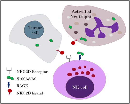
to elucidate these effects but additional binding of NKG2D, an NK cell activating receptor, to an NKG2D-ligand was required. The authors concluded that RAGE/calprotectin has an enhancing or tuning effect on NKG2D induced NK cell activation.

This stimulating paper caused an animated discussion by our Journal Club group focussing on important criteria required for choosing suitable mouse strains and cell lines for a specific model.  We were intrigued by this novel finding and are awaiting the future work by Narumi et al, as they intend to investigate the role calprotectin production by neutrophils, for delivering NK cells activating signals to impede tumor progression.

Read the full article at: http://www.jimmunol.org/content/194/11/5539.full

CD8+ T cells follow neutrophil snail trails in virus-infected airways

Written by Caroline Anderson

Edited by John MacKey

The focus of our journal club this week was a recent paper from Science describing a novel mechanism by which neutrophils deposit chemokine-enriched way-markers to guide CD8+ T cells to the airways during influenza virus infection. (read the full article here http://www.sciencemag.org/content/349/6252/aaa4352.full).


snail-trail-best-jpg
Photo: Krstnn Hrmnsn, Creative Commons License

Whilst the role of chemokines in attracting effector immune cells to sites of immune insult is well established, how localized chemokine gradients are produced and maintained is incompletely understood. Likewise, the molecular mechanisms linking neutrophils and T cell recruitment in viral infections is unclear. Lim et al therefore set out to investigate the hypothesis that neutrophils are responsible for generating chemokine gradients that guide influenza-specific CD8+ T cells in the airways.

The authors first demonstrate that neutrophil depletion in a mouse model of influenza virus infection leads to impaired CD8+ T cell recruitment, localization and function in virus-infected trachea sections. Specifically they observed reduced numbers of both total and virus-specific CD8+ T cells, which lacked motility and remained distal to the epithelium. This dampened T cell response consequently led to a significant increase in viral load, demonstrating the importance of neutrophils in eliciting efficient T cell-mediated viral clearance. Using chemokine arrays and migration assays they were then able to identify CXCL12 as a key neutrophil-derived factor responsible for the recruitment of CD8+ T cells in this model. Importantly, the effects of neutrophil-depletion on CD8+ T cell recruitment were recapitulated using an antagonist against CXCR4, the chemokine receptor recognized by CXCL12. Striking immunofluorescence and scanning electron microscopy images revealed the generation of trails by crawling neutrophils, which were shown to contain CXCL12 and to persist in the trachea for up to 8 days post-viral infection. Finally, time-lapse in vivo two-photon microscopy images showed the deposition of CXCL12+ trails by neutrophils in the mouse ear, which appear to be followed by migrating CD8+ T cells. CXCL12 in neutrophil trails therefore appears to be critical for the effective recruitment and function of influenza-specific CD8+ T cells.

The journal club discussion centered on the exact timeline of neutrophil trail generation and the subsequent recruitment of the CD8+ T cells in vivo. It is clear that the neutrophil trails persist in the trachea for up to 8 days post-infection, yet it would be interesting to map trail deposition by individual neutrophils and to directly relate this to corresponding T cells over the course of the viral infection. In addition, we discussed the potential involvement of other chemokines and factors in this process, as well as the composition of the CXCL12-enriched deposits. We were also curious as to whether the same phenomenon occurred in response to influenza virus infection in other tissues, particularly in the lung, and in response to other viruses or inflammatory insults.

Influenza Promotes Collagen Deposition via αvβ6 Integrin-mediated Transforming Growth Factor β Activation

Journal club with Gisli Jenkins from 21/9/15

Written by David Salman, Richard Toshner and Joana Alçada

Edited by Sarah Allden

We recently welcomed Dr Gisli Jenkins, Clinical Associate Professor & Reader in Pulmonary Biology from the University of Nottingham to the IRD Journal Club.

His recent publication “Influenza Promotes Collagen Deposition via αvβ6 Integrin-mediated Transforming Growth Factor β Activation” served as a starting point to discuss various aspects of research in lung fibrosis. The paper explores the relationship between influenza and idiopathic pulmonary fibrosis via transforming growth factor β (TGF-β), a cytokine involved in development, inflammation and wound repair.

The authors hypothesised that influenza-induced epithelial cell damage would stimulate toll-like receptor 3 (TLR3) activity, a receptor that recognizes by-products from viruses such as influenza. This in turn may lead to activation of an epithelial specific cell surface molecule called integrin αvβ6, which would then turn on TGF-β signalling.

The results first indicate that influenza virus activates TGF-β in airway epithelial cells. Secondly, mutation of the epithelial cells with a non-functioning TLR3 protein inhibited this response, suggesting that the process is mediated through TLR3 signalling. Finally, blocking integrin αvβ6 with an antibody reduced influenza-induced epithelial cell death. Influenza was also shown to increase deposition of matrix materials, which are correlated with pulmonary fibrosis, throughout the lungs of infected mice. In addition, levels of integrin αvβ6 expression were increased in those mice. Conversely, blocking the expression of this integrin with antibody inhibited the development of a fibrosis-like picture in the lungs. Therefore, the Jenkins group have elegantly demonstrated a link between influenza infection and the mechanisms of pulmonary fibrosis development, through integrin αvβ6-mediated TGF- β and TLR3 signalling.

Print

The discussion Dr Jenkins kindly participated in was a wide ranging one, touching upon not just his own group’s research record, but also a more global view of translational research, with particular reference to idiopathic pulmonary fibrosis. He touched upon some of the molecules currently at the phase 2 trial stage for IPF, such as anti IL-13 and anti IL-4, as well as his thoughts upon the possible future directions that research in this area will take.

See Dr Jenkins’ full paper here http://www.jbc.org/content/289/51/35246.long